Biological Sciences
Oncogenic Viruses
Oncogenic viruses are viruses that have the potential to cause cancer in their host cells. They can induce cancer by integrating their genetic material into the host cell's DNA, leading to uncontrolled cell growth and tumor formation. Examples of oncogenic viruses include human papillomavirus (HPV), Epstein-Barr virus (EBV), and hepatitis B virus (HBV).
Written by Perlego with AI-assistance
Related key terms
7 Key excerpts on "Oncogenic Viruses"
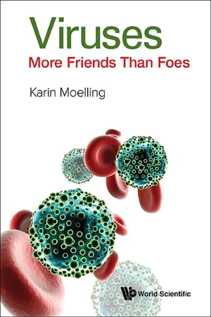 eBook - ePub
- Karin Moelling(Author)
- 2016(Publication Date)
- WSPC(Publisher)
Especially the retroviruses have taught researchers how viruses can contribute to tumors. Such retroviruses are therefore also designated as tumor viruses. They carry cancer genes or oncogenes. Consequently, many retrovirologists think of themselves as cancer researchers — as indeed I do. Many of these viruses are laboratory strains and rarely occur in humans, and, if so, then only in combinations with other factors, which then all together cause cancer — often after decades. However, they can cause cancer in animals, mice, cats, cows, even in plants, and also in monkeys.About 100 viral oncogenes are known. They all are characterized principally by their ability to stimulate cellular growth. Those that do not induce growth advantages, or even suppress growth, do not manifest themselves so easily and are often overlooked. Tumor viruses can lead to small piles of cells in the culture dish, “mini-tumors” designated as foci. They can be big enough to be seen by the naked eye. Researchers picked the biggest foci and thereby selected the viruses with the strongest growth-promoting oncogenes. In animals, viruses with the “best” oncogenes caused the biggest tumors. These viruses helped us researchers to select for the most dangerous cancer genes, now — 40 years or so later — often used by industry as the best targets for anti-cancer therapies which indeed led to today’s most efficient drugs. How test tube oncogenes play a role in human cancers requires explanations, please wait.Normal cells stop growing as soon as they touch their neighboring cells. This phenomenon is well known to everybody who works in the kitchen, where a cut in a finger will be healed by growing cells, which stop dividing as soon as they touch their neighbor cells; this phenomenon is called contact inhibition. This stop signal is missing in tumor cells, which keep growing on top of each other — an important aspect of tumor cell growth.There was a long search to understand where the oncogenes of tumor viruses came from: from the inside or the outside of a cell. To answer that, researchers first had to learn about gene exchange between viruses and cells, Horizontal Gene Transfer (HGT). This is a process whereby genes are transferred in both directions: genes from the inside of the cell get out by means of viruses and the viruses can transfer them back into the inside of new cells by infection. The newly acquired viral genes can be deposited by integration into the cell’s genome. Thus, gene transfer proceeds in both directions. The genes picked up by the viruses are random cellular genes — initially. When we, the experimenters, selected for fast cellular or tumor growth, we ended up with viruses that have special genes, ones that confer upon the cells the greatest advantages in growth — a hallmark of cancer. An additional phenomenon plays a role. The high mutation frequency of viruses contributed further to the most effective growth-promoting carcinogenic genes. The mutations arise during virus replication through the reverse transcriptase, which is highly error-prone, “sloppy”. During each replication, about 10 mutations occur within 10,000 nucleotides, the size of a retrovirus. This high error rate makes the RNA-containing viruses so inventive. Mutagenesis is part of the survival strategy of viruses. Mutations have increased the oncogenicity of the oncogenes. Cells with DNA genomes such as our own cells have developed proofreading mechanisms that largely prevent mutations. That is good for us, because otherwise we would have a higher chance of getting cancer. The cellular genes are known by the abbreviation c-onc (or proto-onc) and after mutations by the virus they are called viral oncogenes v-onc (or viral-onc) — which are the “better”, because stronger, oncogenes. Thus, oncogenes originate from the inside of the cell, but the virus modifies them. eBook - ePub
- Jane Flint, Vincent R. Racaniello, Glenn F. Rall, Theodora Hatziioannou, Anna Marie Skalka(Authors)
- 2020(Publication Date)
- ASM Press(Publisher)
Infection of susceptible cells in culture by members of this family can result in immortalization or induction of typical transformed phenotypes. The large sizes of their genomes initially presented a major impediment to analysis of the transforming properties of these viruses. It is now clear that herpesviral gene products generally alter cell growth and proliferation by mechanisms related to those responsible for transformation by the smaller DNA viruses or retroviruses. However, the genomes of some of these large DNA viruses also encode micro-RNAs (miRNAs) that contribute to transformation (described in Volume I, Chapter 8). Recent Identification of Oncogenic Viruses Oncogenic Viruses associated with human disease continue to be isolated with some regularity. One discovered in 1994 was a previously unknown member of the family Herpesviridae, human herpesvirus 8, which was isolated from tumor cells of patients with Kaposi’s sarcoma. Its genome, like those of transducing retroviruses, contains homologs of cellular proto-oncogenes. More recently (in 2008), a polyomavirus associated with a rare form of skin cancer was discovered (Box 6.6). Perhaps an even greater surprise was the realization that RNA viruses other than retroviruses can be associated with cancer: hepatitis C virus, a (+) strand RNA virus belonging to the family Flaviviridae, is associated with a high risk for hepatocellular carcinoma, although as we shall see, the mechanism by which this virus promotes tumors is quite distinct. Common Properties of Oncogenic Viruses Although they are members of different families (Table 6.1), the majority of Oncogenic Viruses share several general features. In all cases that have been analyzed, transformation is observed to be a single-hit process (defined in Volume I, Chapter 2), in the sense that infection of a susceptible cell with a single virus particle is sufficient to cause transformation eBook - ePub
Principles of Virology, Volume 2
Pathogenesis and Control
- S. Jane Flint, Vincent R. Racaniello, Glenn F. Rall, Theodora Hatziioannou, Anna Marie Skalka(Authors)
- 2020(Publication Date)
- ASM Press(Publisher)
The second class, nontransducing oncogenic retroviruses, are less carcinogenic agents. Not all animals infected with these viruses develop tumors, which appear only weeks or months after infection. In the late 1980s, a third type of oncogenic retrovirus that caused tumorigenesis very rarely months or even years after infection was identified in humans. This group comprises two lentiviruses, human T-cell lymphotropic virus type 1 and 2. Infection by each group of oncogenic retroviruses induces tumors by a distinct mechanism. As their name implies, the genomes of transducing retroviruses contain cellular genes that become oncogenes when expressed in the viral context. The virally transduced versions of these cellular genes are called v-oncogenes, and their cellular counterparts, which are not normally transforming, c-oncogenes or proto-oncogenes. The genomes of the nontransducing retroviruses do not encode cell-derived oncogenes. Rather, the transcription of proto-oncogenes is activated inappropriately as a consequence of the nearby integration of a provirus in the host cell genome. In either situation, the oncogene products ordinarily play no role in the reproductive cycle of the retroviruses themselves. With the notable exception of the reproductive cycle of certain epsilonretroviruses (Box 6.5), the oncogenic potential of retroviruses is an accident of their infectious cycles. Nevertheless, the study of v-oncogenes and proto-oncogenes that are affected by retroviruses has been of great importance in advancing our understanding of the origins of cancer- eBook - ePub
Cancer Chemotherapy
Basic Science to the Clinic
- Gary S. Goldberg, Rachel Airley(Authors)
- 2020(Publication Date)
- Wiley-Blackwell(Publisher)
Colorectal, endometrial, and liver cancer, glioblastoma, melanoma HER2 Epidermal growth factor family receptor tyrosine protein kinase Breast, ovarian, gastric, and prostate cancer KIT Receptor tyrosine protein kinase Small cell lung cancer, gastrointestinal stromal tumor, malignant melanoma, acute myeloid leukemia MYC Nuclear transcription factor Burkitt' lymphoma, multiple myeloma, neuroblastoma, breast cancer, melanoma NOTCH1 Transmembrane receptor, gene transactivation T‐cell acute lymphoblastic leukemia and lymphoma, breast cancer, melanoma PI3K Phosphatidylinositol lipid kinase Breast, endometrial, liver, and colorectal cancer RAS GTPase signal transducer Pancreatic adenocarcinoma, hematopoietic and lymphoid cancer, colorectal cancer, melanoma WNT Growth signaling factor Colorectal, breast, and ovarian cancer4.3 Viral Oncogenes
Oncogenic mutations are often of biological origin, taking place when certain tumorigenic viruses integrate into the host genome. Human tumor viruses may be RNA viruses, including retroviruses and flaviviruses, or DNA viruses, including the herpes, papilloma, and polyoma viruses. These are often associated with certain cancer types as shown in Table 4.2 .Viral oncogenesis may follow infection with RNA or DNA viruses. RNA viruses infect cells and use cellular machinery to undergo reverse transcription to DNA, forming the DNA provirus, which becomes incorporated into the host genome. The DNA provirus can act in two ways by: (i) introducing its own viral oncogene, or (ii) carrying out insertional mutagenesis where viral regulatory sequences augment the expression of host cellular oncogenes.The viral Src gene can trigger neoplastic transformation of infected cells, as described above. Viral homologs for other oncogenes also exist to activate a shared canonical mitogenic pathway. These include the viral Ras GTPases, as well as the v‐Fos and v‐Jun nuclear proteins that form the AP1 transcription factor, as shown in Figures 4.1 and 14.1  eBook - ePub
- Peter G. Shields(Author)
- 2005(Publication Date)
- CRC Press(Publisher)
4 ) noted common characteristics for the epidemiology of virus-associated cancers:- The long induction period between initial infection and the onset of cancer.
- Most candidate viruses are ubiquitous but cancer incidence is rare.
- The initial infection is often subclinical and the time of infection is rarely known.
- Most viral-related cancers require cofactors.
- The causes of cancer may vary by age and by geographic area.
- Different viral strains may have different oncogenic potentials.
- The host factors, especially age of infection, genetic characteristics, and immune status, play a critical role in susceptibility to cancer.
- A virus may play a role at different points in a complex, multistage process of pathogenesis by altering the host’s immune system or by causing a variety of events at the molecular level.
- Many human cancers cannot be reproduced in experimental animals with the putative virus.
- A virus-induced cancer could have the same histologic features with cancers caused by a toxin, chemical, altered gene, or other causal factors.
- The geographic distribution of virus infection is similar to that of the associated tumor when adjusted for the age of infection and the presence of cofactors.
- Viral markers (antibody titers or antigenemia) are higher in cases than in matched controls in the same geographic setting, as shown in case-control studies.
- The viral marker precedes the tumor and a significantly higher incidence of the tumor follows in persons with the marker than in those without it.
 eBook - ePub
Infectious Causes of Cancer
A Guide for Nurses and Healthcare Professionals
- Kenneth Campbell(Author)
- 2011(Publication Date)
- Wiley(Publisher)
In early 2010, the journal Nature published a genome catalogue for the human gut flora 3. Given that microbes have viruses of their own, not considering those infecting our own cells, it is probably impossible to even estimate the amount of viral DNA contained by an average person’s body. Persing has pointed out that 'In purely reductionist terms, infectious diseases can be viewed as horizontally acquired genetic disorders in which exogenously acquired nucleic acids of a pathogen interact chromosomally, episomally, or extracellularly with those of the host to disrupt normal cellular processes or to produce inflammation' 4. It is unsurprising that the disruption of host cell behaviour is capable of giving rise to malignant tumours. Oncogenes and tumour suppressor genes A complex network of regulatory genes controls cell division and the integrity of the genome; cells are programmed to repair errors in DNA copying, and if this fails they are programmed to self-destruct. Unregulated cell division is a threat to the whole organism so normal cells can only divide in response to an external signal - they cannot autonomously 'decide' to divide. Oncogenes drive cell growth and division, whereas tumour suppressor genes restrain this process; they are comparable to a car’s accelerator and brakes. A single (rogue) oncogene can drive cell division; multiple tumour suppressor genes must fail before control is lost, just as most cars have multiple braking systems but a single accelerator. Oncogenes are positive regulators of cell growth, initially recognized as elements of the genome of RNA tumour viruses. The common functional characteristic of oncogenes is that they drive a process required for 'tumour initiation, progression, invasion or metastasis' 5. Stéhelin and his colleagues reported in 1976 that the normal avian genome contains sequences related to the transforming gene(s) of avian sarcoma viruses 6 eBook - ePub
- Antoni Horst(Author)
- 2018(Publication Date)
- CRC Press(Publisher)
The discovery of naturally existing factors inhibiting proliferation of cells creates new bases for understanding the neoplastic processes that carcinogenesis may generate not only by the action of various carcinogens but also because of the loss of the inhibitory factors. In this aspect, particularly important seem to be the emerging possibilities of cancer therapy with such factors as tumor necrosis factor (TNF), transforming growth factor β (TGF-β), and interferons. For successful therapy of cancer with these factors, further investigations are needed, but in the cheerless situation of contemporary cancer therapy, this seems to be the first gleam of hope.X. THE ACTION OF CELLULAR ONCOGENESAt present, about 30 oncogens originating from the cellular genome, and 10 or more present in the genomes of DNA tumor viruses are known. These 40 different oncogenes exhibit various functional properties that can be classified into smaller groups. On the basis of the nuclear or cytoplasmic localization of their gene products (not genes), we distinguish nuclear viral oncogens (SV40 large T-oncogene, polyoma large T-oncogene, and adenovirus E1A) and nuclear cellular oncogenes (myc, myb, N-myc, p53, ski and fos) and cytoplasmic viral oncogenes (polyoma middle T-oncogene) and cytoplasmic cellular oncogenes (ras, src, erbB, neu, ros, fms, fes/fps, yes, mil/raf, mos, and abl).27 The most important feature of all oncogenes is their immortalizing ability (the ability to convert a tissue culture of limited growth to a culture that can be passaged without limits). The nuclear oncogenes (myc, N-myc, myb, and E1A) exhibit some structural homology, whereas some others (p53, polyoma large T-oncogene, and SV large T-oncogene) exhibit some functional similarities. Among the cytoplasmic oncogenes, only ras, src, and middle T-oncogene of polyoma have been studied in detail. The cytoplasmic oncogenes generally exhibit weak immortalizing activity. Nuclear oncogenes collaborate with cytoplasmic oncogenes in malignant transformation of normal cells. Nuclear, but not particularly cytoplasmic oncogenes are able to induce full transformation of cells in culture. An exception is presented by the SV40 large T-oncogene (sis oncogene) which by its single action is able to induce nuclear functions such as immortalization, and cytoplasmic functions such as anchorage independence. It is of interest that the large T-antigen may be found both in the nucleus and at the plasma membrane.27

Learn about this page
Index pages curate the most relevant extracts from our library of academic textbooks. They’ve been created using an in-house natural language model (NLM), each adding context and meaning to key research topics.