Biological Sciences
The Heart
The heart is a muscular organ that pumps blood throughout the body, supplying oxygen and nutrients to the tissues and removing waste products. It is a vital component of the circulatory system and is responsible for maintaining the body's overall function. The heart consists of four chambers and is regulated by electrical impulses to ensure proper blood flow.
Written by Perlego with AI-assistance
Related key terms
6 Key excerpts on "The Heart"
 eBook - ePub
- Britannica Educational Publishing, Kara Rogers(Authors)
- 2010(Publication Date)
- Britannica Educational Publishing(Publisher)
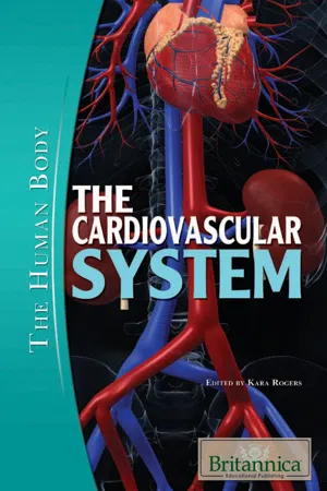
CHAPTER 1THE HUMAN HEARTT he heart is one of the most vital organs in the human body. Its function is to circulate the blood by acting as a pump. With each heartbeat, blood is pushed into the arteries and through the veins. It then courses around the body in a one-way circuit so that it eventually returns to The Heart to repeat the process. Driving this constant movement of the blood are the perpetual rhythmic contractions of The Heart muscle. This defining characteristic of The Heart underlies the body’s ability to routinely and reliably deliver oxygen and nutrients to organs, tissues, and cells.The human heart in situ . Encyclopædia Britannica, Inc.With the exception of some invertebrates, The Heart is an anatomical feature common to members of the animal kingdom. However, the shape and complexity of The Heart varies greatly among the different groups of animals. It may be a straight tube, as in spiders and annelid worms, or a somewhat more elaborate structure with one or more receiving chambers (atria) and a main pumping chamber (ventricle), as in mollusks. In fishes The Heart is a folded tube, with three or four enlarged areas that correspond to the chambers in the mammalian heart. In animals with lungs—amphibians, reptiles, birds, and mammals—The Heart shows various stages of evolution from a single to a double pump that circulates blood (1) to the lungs and (2) to the body as a whole.The human adult heart is normally slightly larger than a clenched fist with average dimensions of about 13 × 9 × 6 cm (5 × 3.5 × 2.5 inches) and weighing approximately 300 grams (10.5 ounces). It is cone-shaped, with the broad base directed upward and to the right and the apex pointing downward and to the left. It is located in the chest (thoracic) cavity behind the breastbone (sternum), in front of the windpipe (trachea), the esophagus, and the descending aorta, between the lungs, and above the diaphragm. About two-thirds of The Heart lies to the left of the midline. eBook - ePub
- Ian Lyons(Author)
- 2011(Publication Date)
- Wiley-Blackwell(Publisher)
6 Cardiovascular systemThe cardiovascular system transports nutrients to tissues, removes waste to be processed, and carries hormones and other signals throughout the body. It is made up of three components: The Heart, the vasculature and the blood.Contraction of The Heart forces blood from its chambers into the pulmonary arteries and the aorta, to be carried around the systemic circulation. Blood flows from The Heart through the arteries to the capillaries, the narrow vessels where diffusion is possible to allow movement of molecules between the blood and tissues. After this, blood drains back to larger veins before it enters the other side of The Heart to be pumped around the pulmonary circulation. The distribution of the blood varies with the need of the tissues themselves, and is achieved through a series of regulatory mechanisms.Given the important function that the cardiovascular system performs, it is not surprising that many pathologies affect it. In particular, there is a potential for fatty deposits to build up, leading to the development of atherosclerotic lesions; these cause narrowing of the vessels and restricted blood flow.Functional anatomy of The Heart External anatomy of The HeartThe external anatomy of The Heart and its relations are covered in Chapter 14. However, a few structures of particular functional importance are covered below. The Heart is located in the mediastinum, enclosed within a fibrous sac – the pericardium. It is connected to the systemic circulation by the aorta and the vena cava, and to the pulmonary circulation eBook - ePub
- John Knight, Yamni Nigam, Jayne Cutter(Authors)
- 2020(Publication Date)
- Learning Matters(Publisher)
Cardiovascular disease such as that which led to George’s heart attack is a major cause of death and disability throughout the world. Nurses will spend a great deal of their time caring for patients with a variety of both heart and blood vessel disease. Many of the routine physiological measurements carried out by nurses such as recording heart rate, blood pressure and blood oxygen saturation provide snapshots of the patient’s current cardiovascular status.This chapter will begin by describing the major functions of the cardiovascular system before examining the structure of The Heart and its associated structures. Once you have an understanding of the anatomy of The Heart, we will explore how The Heart functions as a mechanical pump and examine the control mechanisms that ensure its optimal function. The second section of the chapter will examine the structure of blood vessels and explain how the cardiovascular system maintains cardiac output to ensure that blood pressure and tissue perfusion are adequately maintained. To reinforce the key points, we will explore the nature of some of the cardiovascular diseases that nurses routinely encounter in clinical practice.Overview of the cardiovascular system
The cardiovascular system (Figure 3.1 , Table 3.1 ) consists of The Heart (cardio) and the blood vessels (vascular). It is primarily a pumped system reliant on both the efficient coordinated contractions of The Heart and a system of vessels which act as conduits through which the blood is circulated.Heart structure and function
Position of heart within the thorax
The Heart has a relatively central position within the thoracic cavity (Figure 3.1 eBook - ePub
- Jeremy P. T. Ward, Roger W. A. Linden(Authors)
- 2017(Publication Date)
- Wiley-Blackwell(Publisher)
Part 3 The cardiovascular systemChapters- 19: Introduction to the cardiovascular system
- 20: The Heart
- 21: The cardiac cycle
- 22: Initiation of The Heart beat and excitation–contraction coupling
- 23: Control of cardiac output and Starling’s law of The Heart
- 24: Blood vessels
- 25: Control of blood pressure and blood volume
- 26: The microcirculation, filtration and lymphatics
- 27: Local control of blood flow and specific circulations
 Passage contains an image
19 Introduction to the cardiovascular system
The cardiovascular system comprises The Heart and blood vessels, and contains ∼5.5 L of blood in a 70 kg man. Its main functions are to distribute O2 and nutrients to tissues, transfer metabolites and CO2 to excretory organs and the lungs, and transport hormones and components of the immune system. It is also important for thermoregulation (Chapter 13). The cardiovascular system is arranged mostly in parallel, i.e. each tissue derives blood directly from the aorta (Figure 19.1). This allows all tissues to receive fully oxygenated blood, and flow can be controlled independently in each tissue against a constant pressure head by altering the resistance of small arteries (i.e. arteriolar constriction or dilatation). The right heart, lungs and left heart are arranged in series. Portal systems are also arranged in series, where blood is used to transport materials directly from one tissue to another, such as the hepatic portal system between digestive organs and the liver. The function of the cardiovascular system is modulated by the autonomic nervous system (Chapter 8).Blood vessels
The vascular system consists of arteries and arterioles that take blood from The Heart to the tissues, thin-walled capillaries that allow the diffusion of gases and metabolites, and venules and veins that return blood to The Heart. The blood pressure, vessel diameter and wall thickness vary throughout the circulation (Figures 19.2 and 19.3). Varying amounts of smooth muscle are contained within the vessel walls, allowing them to constrict and alter their resistance to flow (Chapters 12 and 24). Capillaries contain no smooth muscle. The inner surface of all blood vessels is lined with a thin monolayer of endothelial cells, important for vascular function (Chapter 24). Large arteries are elastic and partially damp out oscillations in pressure produced by the pumping of The Heart; stiff arteries (age, atherosclerosis) result in larger oscillations. Small arteries contain relatively more muscle and are responsible for controlling tissue blood flow. Veins have a larger diameter than equivalent arteries, and provide less resistance. They have thin, distensible walls and contain ∼70% of the total blood volume (Figure 19.3). Large veins are known as capacitance vessels and act as a blood volume reservoir; when required, they can constrict and increase the effective blood volume (Chapter 24). Large veins in the limbs contain one-way valves, so that when muscle activity (e.g. walking) intermittently compresses these veins they act as a pump, and assist the return of blood to The Heart (the muscle pump eBook - ePub
- Erin Odya, Maggie A. Norris(Authors)
- 2017(Publication Date)
- For Dummies(Publisher)
Part 4Exploring the Inner Workings of the Body
IN THIS PART … Look at the anatomical structures of the cardiovascular, respiratory, digestive, urinary, and lymphatic systems. Learn how the blood is moved around and how the body gets things into and out of it. Get familiar with the mechanics of breathing and gas exchange. Make sense of the mechanical and chemical breakdown of food. Understand the importance of the kidneys: urine formation and blood pressure control. Investigate immunity and lymphatic flow. Passage contains an image
Chapter 9 The Cardiovascular System: Getting Your Blood Pumping
IN THIS CHAPTERLooking at your blood and what’s in itDiscovering arteries, veins, and capillariesBreaking down The Heart’s partsFollowing blood along its path through your bodyLooking at some cardiovascular system problemsMore than any other system, the cardiovascular system has contributed strong imagery to people’s daily language. “Heart” is a metaphor for love and for courage. People say they “nearly had a heart attack” to describe an experience of surprise or shock. Abstract but distinctive characteristic qualities are said to be “in one’s blood.” The blood itself runs cold, runs hot, and runs all over the place in informal speech and poetry. Scientifically speaking, emotions are more a matter of hormones than myocardium, and nobody’s blood is any redder or any hotter than anyone else’s. The Heart is neither soft nor hard, but a muscular, fibrous pump, and blood is a complex biological fluid that must be kept moving through its specialized network of vessels. Ready to take a closer look?Getting Substances from Here to There
The functions of the cardiovascular system are all related to transportation. Nearly every substance made or used in the body is transported in the blood: hormones, gases of respiration, products of digestion, metabolic wastes, and immune cells. We discuss these transportation functions in the context of the other organ systems only as they relate specifically to cardiovascular anatomy and physiology.
- Ian Peate(Author)
- 2022(Publication Date)
- Wiley-Blackwell(Publisher)
Part 3 The Heart and vascular systemChapters
- 16 The Heart
- 17 Blood flow through The Heart
- 18 The conducting system
- 19 Nerve supply to The Heart
- 20 The structure of the blood vessels
- 21 The blood pressure
- 22 The lymphatic circulation
Passage contains an image
16 The Heart
The location of The Heart.Figure 16.1Source: Peate I, Wild K & Nair M (eds). Nursing Practice: Knowledge and Care (2014).The walls of The Heart.Figure 16.2Source:Peate I, Wild K & Nair M (eds). Nursing Practice: Knowledge and Care (2014).Cells of the myocardium.Figure 16.3Source: Peate I, Wild K & Nair M (eds). Nursing Practice: Knowledge and Care (2014).Myocardial cells.Figure 16.4Source: Peate I, Wild K & Nair M (eds). Nursing Practice: Knowledge and Care (2014).The heart is part of the cardiovascular system. It weighs approximately 250–390 g in men and 200–275 g in women and is a little larger than the owner’s closed fist, being about 12 cm long and 9 cm wide. It is located in the thoracic cavity (chest) in the mediastinum (between the lungs), behind and to the left of the sternum (breastbone) (Figure 16.1 ). The Heart rests on the diaphragm in the thoracic cavity.Walls of The Heart
Pericardium
A membrane called the pericardium (peri = around) surrounds The Heart. This is referred to as a single sac surrounding The Heart but is in fact made up of two sacs (fibrous pericardium and serous pericardium) that are closely connected to each other (Figure 16.2 ). These two sacs have very different structures.Fibrous pericardium
This is a tough, inelastic layer made up of dense, irregular, connective tissue. Its purpose is to prevent overstretching of The Heart. It also provides protection to The Heart and anchors it in place.Serous pericardium
The serous pericardium is a thinner, more delicate structure which forms a double layer around The Heart. The outer layer is fused to the fibrous pericardium. The visceral pericardium (otherwise known as the epicardium) adheres tightly to the surface of The Heart.
Learn about this page
Index pages curate the most relevant extracts from our library of academic textbooks. They’ve been created using an in-house natural language model (NLM), each adding context and meaning to key research topics.