Physics
Radiotherapy
Radiotherapy is a medical treatment that uses high-energy radiation to target and destroy cancer cells. It is based on the principles of physics, specifically the interaction of radiation with matter. Radiotherapy can be delivered externally using machines like linear accelerators or internally through radioactive implants. The goal is to shrink tumors and prevent cancer from spreading.
Written by Perlego with AI-assistance
Related key terms
10 Key excerpts on "Radiotherapy"
 eBook - ePub
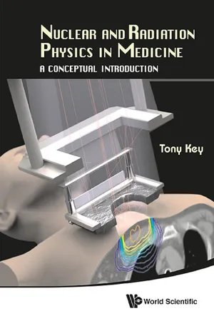
Nuclear and Radiation Physics in Medicine
A Conceptual Introduction
- Tony Key(Author)
- 2013(Publication Date)
- WSPC(Publisher)
Chapter 4Radiation Therapy4.1 IntroductionPrevious chapters have described the use of radiation as a diagnostic tool. However, in its passage through matter, radiation causes ionization that can damage the cells through which it passes. At the low levels of radiation used in diagnosis, the body can usually repair this damage; at higher levels, illness can result. However, since radiation preferentially destroys cancer cells, which grow at a faster rate and repair themselves less well than do healthy cells, radiation has become a powerful tool in the treatment of cancer.In radiation therapy, the intent is to sterilize or kill the tumour cells while causing minimal damage to adjacent healthy tissues. Many different sources of radiation are used. There are two main methods of delivery of the radiation: teletherapy, where the radiation from X-rays, radioisotopes, or external particle beams (electrons, protons, neutrons, or heavy ions) is directed into the body from the outside, and brachytherapy (the root comes from Greek, meaning short distance), in which a radioisotope is placed on or inside the body, close to the site of the cancerous tissue.Section 4.2 discusses the mechanisms whereby biological cells are damaged by radiation. Section 4.3 shows some basic methods used to calculate the required therapeutic doses from external radiation. Section 4.4 discusses beams of radiation from accelerators and from radioisotopes. Brachytherapy, its uses, and related dose calculations appear in section 4.5 .Chapter 5 provides a short overview of the unavoidably deleterious effects of radiation on healthy cells and the contribution of both diagnostic and therapeutic radiation to the environmental radiation background.4.2 The interaction of radiation with biological cellsX-rays and the emissions from radioactive isotopes (alpha, beta, or gamma rays) ionize the cells through which they pass. (Ionization is the process whereby electrons are removed from an atom or molecule to form an ion or a charged molecule). Alpha particles (nuclei of He atoms, see §3.2.2 ), by virtue of their greater mass and double charge, produce a greater density of ionization than beta particles (electrons or positrons). As a consequence, alpha particles are much more weakly penetrating than beta particles, which in turn are less penetrating than X-ray or gamma ray photons at a given energy. Alpha particles of typical energy are easily stopped by, e.g. a sheet of paper, or human skin; however if they are ingested, their high ionizing ability causes cellular damage to the lungs, which is the main reason smoking causes cancer (see §5.1.2 ). Photons of X- or gamma rays are called indirectly ionizing radiation since they do not directly cause biological damage, but produce energetic electrons via the processes described in §2.3 eBook - ePub
- Mark Strikman, Kevork Spartalian, Milton W. Cole(Authors)
- 2014(Publication Date)
- Princeton University Press(Publisher)
CHAPTER 8
Applications in Treatment
8.1 OVERVIEW
Medical treatment of patients has diverse components, including the use of medicines or physical therapy and the application of many techniques derived from the discoveries of modern physics. In this chapter, we describe the last of these categories of treatment. Just as modern physics has enabled us to explore and analyze the previously hidden interior of patients, these techniques provide means of repairing, or even replacing, diseased tissues and organs located deep within the patient. In addition, the laser provides a means of treating problems situated at, or near, the surface of a patient, as in LASIK eye surgery. A common ingredient in all these methods is the need for a precise focus of the treatment, since otherwise significant harm may result. Evidently, those caregivers providing the treatment must work closely with those providing the diagnosis.8.2 TREATMENT WITH RADIATION
In section 5.2 , we discussed the biological effects of radiation. Because radiation can kill cells, it can be used for the treatment of various tumors. The goal of radiation therapy is to deliver an optimal dose of radiation to all parts of the tumor while minimizing the radiation impinging on the rest of the body, especially to radiosensitive tissues like intestines. Three strategies are used to reach this goal: brachytherapy, radioimmunotherapy, and external-beam Radiotherapy.8.2.1 Brachytherapy
The name brachytherapy has its origin in the Greek word brachys, meaning “short.” In brachytherapy, radioactive material is first encapsulated and then placed inside or in close proximity to the tumor, either permanently (low-dose rate) or for a short time (high-dose rate).One example of the low-dose treatment involves treating prostate cancer by permanently implanting radioactive “seeds,” that is, small particles, with positioning guided by ultrasound or MRI. An advantage of brachytherapy is that one can reduce significantly the dose to the healthy tissues by choosing isotopes that emit photons or electrons and have a sufficiently short half-life. Commonly used photon emitters are 192 Ir, 137 Cs, 125 I and 103 Pd. Common electron emitters are 90 Sr and 90 Y. As might be seen in an extrapolation of figure 3.18 to larger A and Z, these two isotopes lie on the neutron-rich side of the nuclear stability curve, so they undergo beta decay. Since photons and electrons have a short mean free path, they are predominantly absorbed close to the emission source. The rate of their interaction, as a function of L, the distance from the source, is proportional to Aμ exp(-μL)/L2 . Here A is the activity of the source, and μ is the absorption coefficient of the emitted particles (see section 4.2 eBook - ePub
Clinical Oncology
Basic Principles and Practice
- Peter Hoskin(Author)
- 2020(Publication Date)
- CRC Press(Publisher)
5 Principles of RadiotherapyRadiotherapy is the use of ionizing radiation to treat disease. The radiation used is either produced by specialist equipment designed for treating patients or by using a specific radioisotope that decays in a set manner to produce radiation.Types of radiationAlpha ( α ) : A heavy charged particle that has classically been difficult to harness in the treatment of patients. Radium 223 is an alpha emitter used in the treatment of metastatic prostate cancer.Beta ( β ) : An electron produced by the decay of certain radioisotopes or by specialist Radiotherapy equipment. Electrons are an important part of therapeutic radiation treatment.Gamma ( γ ) : A beam of radiation, part of the electromagnetic spectrum. Gamma rays (or x-rays) are produced by the decay of certain radioisotopes and from specialist radiation equipment. Gamma radiation is the main type of radiation used to treat cancer patients (and in diagnostic CT units).Proton : Protons are available in an increasing number of centres around the world and the first centre in the UK became operational in 2018. Their advantage is the production of a highly localized, high-energy peak of energy deposition (Bragg peak ), which, by manipulation of the beam energy and by the use of absorbing materials, can be focused to a defined position in a patient. Biologically they are similar in action to x-rays. They have been used particularly for tumours in inaccessible sites such as the back of the eye, base of the skull and childhood brain and spinal tumours. Research is ongoing to see if they have any advantage for other disease sites.Neutron eBook - ePub
The Molecular Biology of Cancer
A Bridge from Bench to Bedside
- Stella Pelengaris, Michael Khan(Authors)
- 2013(Publication Date)
- Wiley-Blackwell(Publisher)
18 Treatment of Cancer: Chemotherapy and Radiotherapy Anne L. Thomas, J.P. Sage, and William P. Steward University of Leicester, UK Key Points More patients than ever before are being cured of their cancer or are having ongoing treatment to allow them to live with their cancer with good quality of life. Major advances have been made in the delivery of Radiotherapy to minimize the damage to normal tissues. The increased understanding of tumor biology has resulted in many biologically targeted agents being available in the clinic. For a number of the common tumor types, genetic testing of the tumors of patients is now standard practice to define which targeted therapies they should receive. Consideration regarding the future cost of cancer therapy needs to be made to ensure that the complex therapies developed are not prohibitively expensive and therefore not financially viable. Introduction Chemotherapeutic agents exert their effect by killing cells that are rapidly dividing. The agents are therefore not tumor cell specific and cause their toxic effect by killing dividing normal cells (e.g. hair follicle cells and gastrointestinal (GI) mucosa). In recent years, our understanding of the molecular pathways controlling the growth of both normal and tumor cells has improved significantly. By exploiting the differences between normal and malignant cells, we can target pathways and receptors unique to the cancer cells, thus avoiding the indiscriminate universal killing of dividing cells of conventional cytotoxics. We are therefore entering the era of biologically targeted therapies in cancer treatment (Chapter 16). Radiotherapy is the application of ionizing radiation to treat disease. Ionizing radiation is electro-magnetic radiation and elementary particles, which deposit energy in materials through the processes of excitation and ionization events. The forms of ionizing radiation in common use are photon beams (X-rays and gamma rays) and electrons (β particles) eBook - ePub
- John E. Niederhuber, James O. Armitage, James H Doroshow, Michael B. Kastan, Joel E. Tepper(Authors)
- 2019(Publication Date)
- Elsevier(Publisher)
- • Image-guided radiation therapy uses real-time and/or daily imaging to ensure that the tumor is positioned such that the radiation beams are precisely delivered to the appropriate location within the patient.
Other Modalities in Radiation- • Brachytherapy, the placement of radioactive sources immediately adjacent to the tumor, delivers extremely high-dose radiation to tumor tissue with a much lower dose to surrounding normal tissues.
- • Stereotactic radiosurgery and stereotactic body radiation therapy combine high dose-per-fraction irradiation with highly conformal treatment delivery to increase the therapeutic ratio while reducing overall treatment time.
- • Proton therapy has dose distribution advantages over photon therapy, and it may be used to deliver high doses of radiation to tumors in close proximity to sensitive normal structures.
Radiation therapy is one of the three established cancer treatment modalities and is used for the therapy of most types of solid tumors and for selected hematologic malignancies. It is used almost entirely for therapy of malignant disease, although it has a small role in preventing proliferation in selected benign diseases. Radiation therapy is routinely combined with surgery, chemotherapy, or both to improve therapeutic results. It is often used with surgery to destroy microscopic regions of tumor extension and with chemotherapy to destroy more effectively the primary tumor. An understanding of the therapeutic use of ionizing radiation requires a basic comprehension of both the physics of radiation therapy delivery and the biologic effects of the interaction of radiation with matter.Overview of Radiation Physics
The toxic biologic effects of ionizing radiation, although complex, varied, and incompletely understood, form the basis for the use of radiation therapy as a cancer treatment. These biologic effects are initiated when packets of energy are deposited in a volume of tissue and remove electrons from constituent atoms through a process called ionization. eBook - ePub
- Suzanne Amador Kane, Boris A. Gelman(Authors)
- 2020(Publication Date)
- CRC Press(Publisher)
For some regions of the body, radiation is especially effective (e.g., in the treatment of superficial skin cancers). For others the responsiveness of tumor cells can differ little from that of neighboring healthy tissues. The cure rate, and the effectiveness of radiation as a cancer treatment, depends greatly on the type of cancer and specifics of individual cases. Physicians treat cancer through a variety of approaches, including surgical removal, chemotherapy, radiation therapy, immunotherapy, and hormone therapy, several of which may be tried in combination or succession. In planning radiation therapy, physicians called radiation oncologists weigh all the information available to them about a specific case: the type of cancer, its history and location, the likely effectiveness of surgery and chemotherapy, other complicating conditions in the patient, and many other factors. Radiation therapy may be used as a palliative measure to reduce pain or deformation even if a cure is unlikely. Factors that can be controlled include the means of delivering the radiation, the type of radiation, total dose size, fractions into which the total dose is divided, and the time between fractions. The type of particle used and its energy are essential issues in radiation therapy. Since the main goal is to have the tumors absorb energy from radiation, high-LET particles might seem like the obvious choice. However, the very short range of alpha particles (Figure 6.2) prevents them from reaching the entire diseased region. Neutrons have a longer range, but require a nearby nuclear reactor for their production. Even more exotic particles have been tested for radiation therapy, but presently, most therapy is performed with either high-energy photons or beta particles (electrons). Electrons with energies from 4 to 20 MeV have ranges of 1 to 6 cm eBook - ePub
- Jeffrey S. Tobias, Daniel Hochhauser(Authors)
- 2013(Publication Date)
- Wiley-Blackwell(Publisher)
5
Radiotherapy
Sources and production of ionizing radiationRadioactive isotopes Artificial production of X-rays and particlesBiological properties of ionizing radiationTumour sensitivity Fractionation and cell deathResponse of biological tissues to radiationNormal tissues Highly radiosensitive tissues Moderately radiosensitive tissues Less radiosensitive tissuesLate sequelae of radiationCarcinogenesis Teratogenicity MutagenicityTechnical aspects of RadiotherapyRadiotherapy planning and treatment techniquesTreatment prescription Maximum, minimum and modal dose Open (direct) and wedged fields Parallel opposed fields Shrinking field technique Systemic irradiation Immobilization devices Conformal and intensitγ-modulated radiation therapyIntegration of Radiotherapy and chemotherapyA recent report from the Royal College of Radiologists (UK) estimated that, taking cancer patients as a whole and assessing the contributions of differing modalities to cure rates, ‘of those cured, 49% are cured by surgery, 40% by Radiotherapy and 11% by chemotherapy’ [1].Ever since the discovery of X-rays by Roentgen in 1895, attempts have been made not only to understand their physical nature but also to use them both in the biological sciences and in a variety of human illnesses. The development of the X-ray tube rapidly led to clinical applications, first as a diagnostic tool and later for therapy in patients with malignant disease. The discovery of radium by Marie and Pierre Curie in 1898 also resulted in the use of radioactive materials for the approach to cancer, since surgery was the only alternative available at that time. Over the past 80 years our understanding of the physical characteristics, biological effects and clinical roles of ionizing radiation has greatly increased. Important articles and papers are listed under Further reading (pages 75–76). eBook - ePub
- Anders Brahme(Author)
- 2014(Publication Date)
- WSPC(Publisher)
The Radiation Biological Basis of Radiation Therapy 3 Anders Brahme, Panayiotis Mavroidis, and Bengt K. Lind3.1. Introduction
Today surgery and radiation therapy are the major treatment modalities for curing cancer patients. About 20–25% of the diagnosed cancers can be cured by each of these treatment modalities either alone as sole modalities or together in adjuvant treatments. In addition, cytostatic drugs, kinase inhibitors, and other chemotherapeutic agents can help cure about 5–10% of the diagnosed cancers. Thus, over 50% of all cancer patients may be cured and this is also true for that half of the patients who are receiving radiation therapy (cf. Chapter 8 , Fig. 8.40 ).During the past three decades, radiation therapy has gone through a very dramatic development: during the 1970s and 1980s computed tomography (CT) and magnetic resonance imaging (MRI), respectively, have revolutionized the diagnostic phase of radiation therapy by providing truly three dimensional (3D) diagnostic tools of sub-millimeter accuracy. This phase has matured further during the 1990s and has been amplified by an equally important development of radiation therapy equipment allowing truly 3D dose delivery to the tumor. The time between the first introduction of new advanced medical tools and the full realization of the potential benefits in the clinic is often quite long, especially considering the long follow-up periods (5–10 years) required for many cancer sites. Hopefully, during the first decade of the new millennium we will start to see the benefits of these developments in improved local cure and survival as new 3D intensity-modulated treatment modalities are taken into clinical use.After a brief review of the whole Radiotherapy process, the main goals of radiation therapy will be formulated in radiobiological terms. Furthermore, the treatment results of classical and advanced intensity-modulated radiation therapy will be discussed. Finally, the different treatment modalities employed today for radiation therapy as well as the characteristics of the different treatment units will be discussed and the future development of radiation therapy will be indicated. eBook - ePub
- B.H Brown, R.H Smallwood, D.C. Barber, P.V Lawford, D.R Hose(Authors)
- 2017(Publication Date)
- CRC Press(Publisher)
Research into the nature of fundamental particles has resulted in megavoltage x-ray generators that can be used in therapy. The use of ionizing radiation in therapy depends on the fact that tumour cells are more susceptible to radiation damage than normal cells. The radiation used is usually either x-rays (produced by decelerating electrons) or ϒ-rays from radioactive materials. The effects of x- and ϒ-ray photons on tissue are the same, and no distinction will be made between them.The radiation dose which can be delivered to the tumour depends on the source-to-skin distance, how well the radiation penetrates the tissues, and how much radiation is scattered into the treatment area from the tissues outside this area.If a beam of x-rays diverges from a point source, and the medium through which the beam passes does not absorb any energy, the intensity of the beam at any given distance from the source will be governed by the inverse square law. Energy will be removed from the beam when an absorbing medium, such as a person, is placed in the beam. Scattering will cause photons to be diverted from their path and absorption will transfer energy from the photons to the electrons of the absorbing medium. The electrons lose their energy by producing ionization and excitation along their tracks, thus causing radiation damage. The excitation process raises the energy level of an electron in an atom, and the ionization process causes electrons to be ejected from atoms. For incident photons of high energy their energy is dissipated over a distance that may be several millimetres in tissue and all electrons lose their energy and come to rest.Several processes contribute to the absorption, and were considered in detail in section 5.2. In the photoelectric process, all the energy of an incident photon is transferred to an electron which is ejected from the atom. This is the predominant process for low-energy photons in materials of high atomic number, i.e. for diagnostic x-rays in bone or metals. Photoelectric absorption produces the high contrast between bone and soft tissue in diagnostic radiographs. eBook - ePub
- Christopher Griffiths, Jonathan Barker, Tanya Bleiker, Robert Chalmers, Daniel Creamer(Authors)
- 2016(Publication Date)
- Wiley-Blackwell(Publisher)
CHAPTER 24 Principles of Radiotherapy Charles G. Kelly and John Frew Northern Centre for Cancer Care, Freeman Hospital, Newcastle upon Tyne, UKIntroduction
The clinical effects of ionizing radiation on the skin have been known since the discovery of X-rays in 1895 [1, 2]. Initially both benign and malignant skin conditions were irradiated and dose and clinical indications were chosen empirically with little knowledge of the late effects of radiation on skin and subcutaneous tissue. Indications for treating benign disease by irradiation have declined since the advent of topical steroids.It is in the best interest of patients with skin tumours to be seen in a clinic where the expertise of specialists in Radiotherapy and oncology, plastic surgery and micrographic surgery as well as dermatology are present, and this is being achieved with the mandatory development of multidisciplinary team meetings and clinics in the UK as described in improving outcomes guidance [3].Ionizing radiation in the treatment of skin cancer
X-ray photon beams: megavoltage and kilovoltage X-ray therapy
X-rays are part of the electromagnetic spectrum, of shorter wavelength and more energetic than UV light. The most commonly used form of therapeutic radiation is megavoltage Radiotherapy using energies of greater than 1 million electron volts (>1 MeV), delivered by linear accelerators in the treatment of deep-seated tumours. Previously, these beams have usually not been suitable for the treatment of skin cancers because of their greater penetration and skin sparing properties, but that has changed in the last few years, with advances in beam delivery technology, although megavoltage beams are still only used for a small minority of skin cancer patients and in particular clinical situations. X-rays in the kilovoltage range (50–140 kV) are very suitable for treating skin cancer as their maximum energy is deposited on the surface. Fall off subsequently is exponential (Figure 24.1
Learn about this page
Index pages curate the most relevant extracts from our library of academic textbooks. They’ve been created using an in-house natural language model (NLM), each adding context and meaning to key research topics.